The Application of Bone Mineral Density Measurement in Digital Radiography System
Osteoporosis is a metabolic bone disease, in which bone mass density gets lower due to the loss of important bone minerals and calcium. Remaining undiagnosed and untreated, it can lead to serious medical conditions resulting in bone fractures. In the worst scenario, it may raise the chance of mortality due to complications. The Bone Mineral Density (BMD) Test can help, which is conducted to evaluate the density of bone before the condition becomes worse and is prone to fracture and breakage. It often works with the digital radiography system, an important diagnostic technology in today’s clinical practice. The image produced by the digital radiography system is useful in diagnosing osteoporosis, predicting future fracture risk, and evaluating the effectiveness of any osteoporosis treatment. An x-ray image by digital radiography system is a convenient way in the diagnosis of osteoporosis, the prediction of the risk of future bone fracture, and the assessment of the efficacy of osteoporosis treatment.

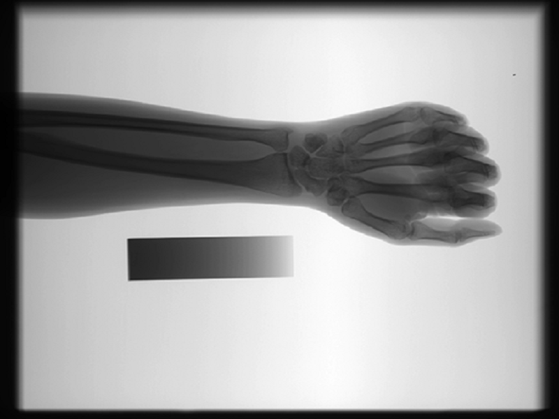
Figure 1. Image produced by digital radiography system for bone mineral density measurement
Challenges in the Bone Density Measurement
In the current clinical practice, the measurement of bone density is achieved via noninvasive BMD methods, among which the single X-ray absorptiometry (SXA) method is widely used. Despite the help of the digital radiography system, to achieve a high accuracy from the result of the bone density measurement could still be challenging when the Ulna and Radius (UR) are not accurately extracted from forearm radiographs for analysis. Hence, automatic segmentation of UR in forearms radiographs is a crucial first step. In the widely used single x-ray absorptiometry (SXA) method in BMD measurement, the accurate extraction of UR from forearm radiographs is a high requirement task due to the differences in forearm between patients and the non-uniform intensity of forearm radiographs.
Research by SONTU for Bone Density Measurement
For efficient evaluation of bone mineral density, we (Shenzhen SONTU Medical Imaging Equipment Co. Ltd) have conducted advanced research based on 142 radiographs and proposed a practical method for automatic segmentation of UR by utilizing the dynamic programming (DP) algorithm, which can be applied to the digital radiography system. This is how our method works:
- First, crop irrelevant regions and downsample the cropped image.
- Second, adjust the direction of the hand to standard. This step is to eliminate the variation due tothe hand direction.
- Third, extract the forearm and hand mask. The intensities in the masked regions are included inthe range [0, 1].
- Fourth, use a bilateral filter [13] to reduce noise and the histogram equalization algorithm to enhance the contrast.
- Then, obtain a standard input image.
- Next, detect four points at the edges of UR diaphysis as the seed points and compute two cost maps (vertical and horizontal) on the preprocessed image.
- Finally, apply the DP algorithm to trace four UR diaphysis segments from four seed pointsand two UR segments to close the UR contours.
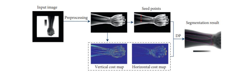
Figure 2. The flowchart of our method
On the basis of our method, the average Dice similarity coefficient reached 0.945. The average mean absolute distance between the contour extracted using this method by the radiologist was only 5.04 pixels. There was no significant difference in the segmentation performance between normal and low-exposure radiographs. The method was validated on 105 forearm x-rays obtained from several hospitals under different imaging conditions. The practicability and operability of this method make it ideal for applying to the digital X-ray system, thereby achieving the accurate extraction of UR in forearms radiographs and generating efficient images. Utilizing our method empowers a more accurate and efficient bone density measurement for diagnosing metabolic and endocrine bone diseases with the digital radiography system.
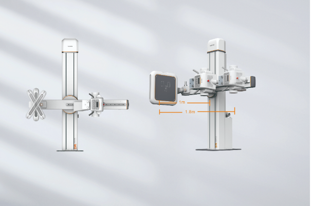
Figure 3. The digital radiography system that can be used for bone mineral density measurement
Digital Radiography System from SONTU Based on the SXA Method Increases the Accuracy of Bone Density Measurement
SONTU Medical Imaging uses the SXA method in the DR system for bone mineral density measurement. The integrated solution, SONTU digital radiography system produces high-quality x-ray image and the dedicated algorithm ensures great accuracy of the measurement, together lays solid foundation for the optimal result of the bone density analysis and helps the cost efficiency from the additional BMD device. Furthermore, SONTU’s digital radiography system is ergonomically designed to better facilitate the advanced clinical practice and provide patient-centered care. SONTU, the professional X-ray machine manufacturer for digital radiography system. Contact us for more details.
References:
- Automatic Segmentation of Ulna and Radius in Forearm Radiographs Available at: https://www.ncbi.nlm.nih.gov/pmc/articles/PMC6374800/